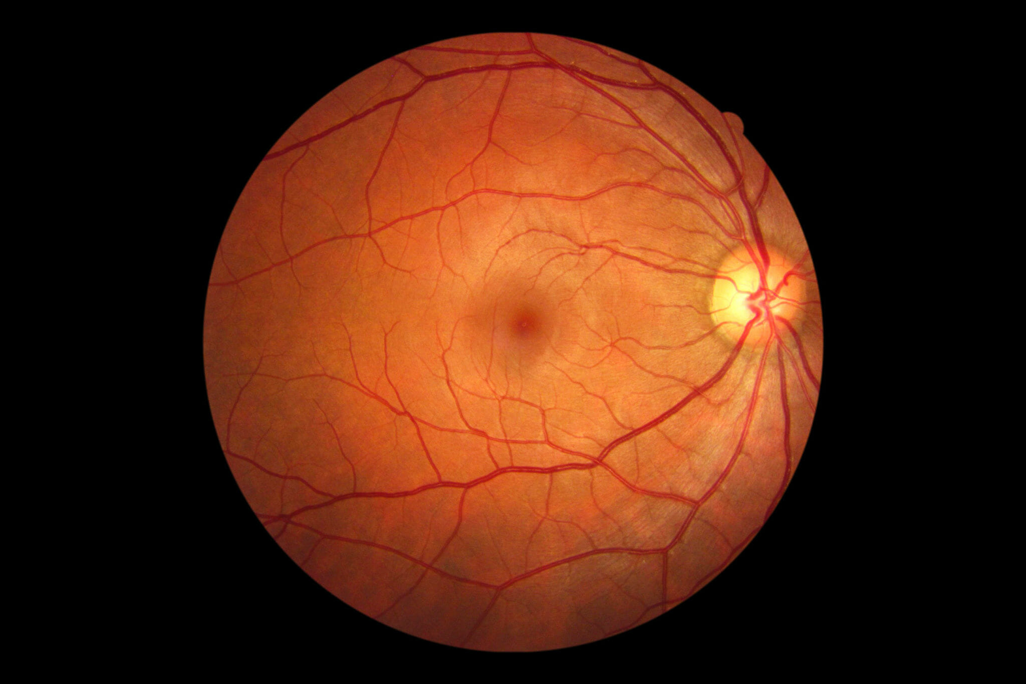

There was no neovascularization anteriorly, but a diffuse VH was seen in the right eye. She was diagnosed with a VH secondary to proliferative diabetic retinopathy (PDR) in the right eye.Īnother patient, a 67-year-old Hispanic male, presented with blurred vision OD for five days accompanied by dark spots and “spider webs.” Visual acuities were 20/100 OD and 20/20 OS. She reported uncontrolled blood glucose levels consistently measuring above 300mg/dL. The patient’s medical history was significant for type 1 diabetes. The left had hemorrhages and intraretinal microvascular abnormalities without neovascularization. The right fundus had preretinal and vitreous hemorrhages (VHs) centrally, intraretinal hemorrhages and neovascularization along the major arcades ( Figure 1 ). Intraocular pressures, pupils and anterior segment findings were normal.

Visual acuities were 20/70 OD and 20/50 OS. Retinal detachment is serious and can cause blindness if not treated quickly.A 35-year-old Hispanic female presented to the ophthalmic emergency department with vision loss OD for one week. When the retina is detached it does not work properly and your vision becomes blurry. After surgery most people can go home after being observed for a short while. Vitrectomy Surgery - is an outpatient surgery and involves the removal and replacement of the vitreous gel from the eye, and attach the retina with the help of lasers to retinal breaks and injecting gas or silicon into the eye to hold the retina in place until it seals. When the tear seals fluid under the retina gets absorbed. The ‘buckle’ holds the retinal tear against the sclera, the tear is then sealed using freezing probe or laser beam. The procedure involves a piece of silicone plastic or sponge being sewn onto the site of a retinal tear to move the sclera towards it. Scleral Buckle – is a surgery performed in the operating room either under local or general anesthetic. The tear is then sealed up in the retina using a freezing probe or laser beam. The bubble is manipulated until it arrives at the detached area.

The eye is numbed with local anthesis and a gas bubble injected into the middle of your eye. Pneumatic Retinopexy - is an outpatient surgery used for certain types of retinal detachments repairs. This type of detachment can be caused by age-related macular degeneration, tumors or inflammatory disorders. Usually related to people who suffer from diabetes or other medical conditions that cause retinal blood vessels to grow.Įxudative – Fluid is found accumulating beneath the retina even though there are no holes or tears. The retina is eventually pulled away from the back of the eye. Tractional – Caused by proliferation of retinal blood vessels, which starts pulling on the retina. The retina detaches, losing its blood supply resulting in a loss of vision. This is the most common type of retinal detachment and caused by a tear that allows fluid to collect underneath the retina. Rhegmatogenous – Caused usually by aging.

Fluid may pass through a retinal tear, lifting the retina off the back of the eye.Ī lot of new gray or black specks floating in your field of vision (floaters)Ī dark shadow or “curtain” on the sides or in the middle of your field of vision Retinal detachment is an eye problem that happens when your retina (a light-sensitive layer of tissue in the back of your eye) is pulled away from its normal position at the back of your eye.


 0 kommentar(er)
